Starting this fall, Catholic Medical Center (CMC) is introducing two new technologies that will allow physicians to get a closer look at the heart. Cardiac PET and cardiac MR will allow cardiologists and radiologists see finer, more detailed images of the heart in patients who may not otherwise get answers from traditional testing.
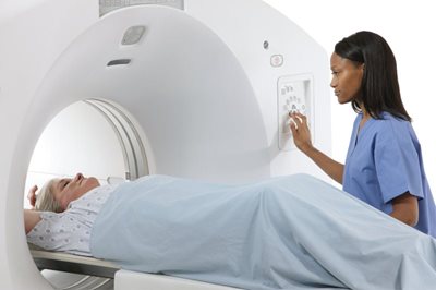
CMC will be the first facility in New Hampshire to offer Cardiac PET. PET stands for Positron Emission Tomography. It’s a lower-radiation test that measures blood flow and captures high-quality images of heart muscle and tissue. Cardiac PET works by using a special camera and a small dose of an isotope, which is administered through an IV. The test helps doctors evaluate the damage done by a heart attack and diagnose coronary artery disease.
“In many instances, a stress test doesn’t give us the information we need to make a solid diagnosis,” says New England Heart & Vascular Institute (NEHVI) cardiologist Ido Preis, MD, FACC. “Sometimes a patient isn’t able to take a stress test. Other times, for one reason or another, the image quality is poor. Cardiac PET produces high-quality images, giving us another tool to deliver the best care to every patient, every time.”
Cardiac MR is an especially helpful tool that uses MRI technology to evaluate heart structure. “It’s the gold standard for studying the structures of the heart,” says Vikas Veeranna, MD, who, along with Peter Shaw, MD, is joining CMC to launch the cardiac MR program.
Dr. Veeranna explains that not all cases of cardiomyopathy (a condition that can lead to heart failure) or heart failure have a clear cause. Certain kinds of cardiomyopathy, for example, can run in families.
“Ten or fifteen years ago, these patients would have been put in a very broad category without a specific diagnosis. They didn’t fit into standard treatment courses, either. Cardiac MR allows us to see very clearly what’s going on in their heart so we can give them an individualized diagnosis and specialized therapy for their needs.”
In the past, these patients may have needed a heart tissue biopsy or gone without a definitive diagnosis. Now, they have a non-invasive, no-radiation option. “It’s a true game changer,” says Dr. Veeranna.
“Imaging technology has advanced significantly, even in the last few years,” says Marcy Rushford, CMC’s director of imaging services. “The result is better treatments and a better experience for patients. Not only do these newer technologies produce more precise images than ever before, they’re often less invasive, quicker, and more comfortable for patients.”
Bringing these technologies to NEHVI is part of CMC’s commitment to be at the forefront of cardiac care. “State-of-the-art imaging is a major driver in how we manage patients,” says NEHVI Executive Medical Director Louis Fink, MD, FACC. “It helps us in our medical management, our surgical expertise, and in our understanding of heart disease.”
That has significant implications for both the treatment of individual patients and for prevention for the population as a whole. Heart disease is still a leading cause of death in the United States and in New Hampshire, even though people are more frequently surviving heart attacks and other cardiovascular events. When doctors are able to dig deeper and better understand the causes of heart disease and heart failure, says Dr. Veeranna, “we can do more to prevent these kinds of episodes from happening.”
Published in the Union Leader's Medical Journal 8/24/19